Li Lab Research

Preventing neurological disease progression through biomarkers, circuits, and neuroengineering interventions.

Our work combines chronic electrophysiology, optical and MR imaging, graph theory, automated event detection, and non-invasive photobiomodulation to understand and prevent epileptogenesis, cognitive decline, and related neurological dysfunction.

Contribution 1

Non-invasive photobiomodulation for prevention-oriented neurology.

The lab develops repetitive transcranial photobiomodulation (PBM) protocols to test whether light-based, non-invasive stimulation can reduce pathological activity before chronic disease progression is established.

Across epilepsy and Alzheimer's disease models with epileptiform activity, this work measures short-term electrophysiological endpoints, including interictal spikes, seizures, spike-ripple events, and high-frequency oscillations, alongside longer-term memory and disease-progression outcomes.

InterventionRepeated wavelength- and site-specific PBM protocols.
ReadoutsSpikes, seizures, HFOs, memory, and network activity.
ModelsmTLE, post-traumatic epilepsy, and AD-related epileptiform activity.

Representative publications

You et al. Preventive effects of transcranial photobiomodulation on epileptogenesis in a kainic acid-induced rat epilepsy model. Experimental Neurology, 2025.

Kang et al. Site- and EEG-frequency-specific effects of 800-nm prefrontal PBM on EEG global network topology. Neurophotonics, 2025.

Wang et al. Photobiomodulation as a potential treatment for Alzheimer's disease. Brain Sciences, 2024.

You et al. Preclinical studies of transcranial photobiomodulation in neurological diseases. Translational Biophotonics, 2020.
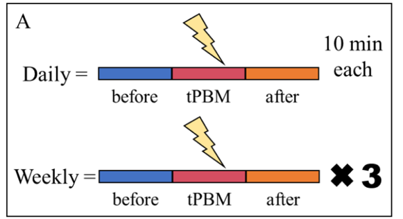
Photobiomodulation experimental timeline
PBM treatment and recording timeline
Brain slice electrophysiology data
Slice and electrophysiology readouts
pHFO network analysis
pHFO-based network analysis
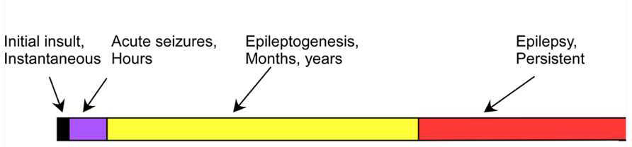
Epileptogenesis timeline
Latent-period epileptogenesis timeline
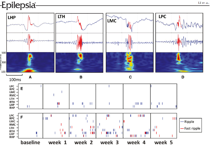
High-frequency oscillation waveform
High-frequency oscillation features
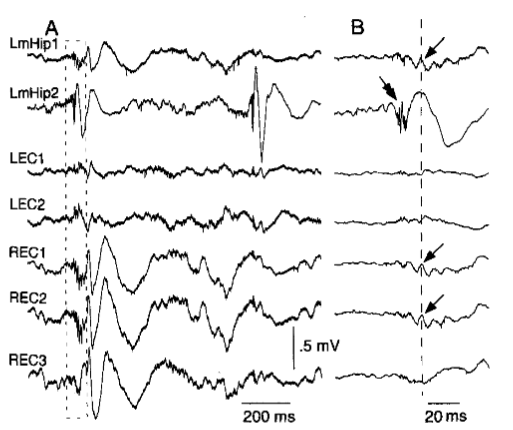
Voltage depth profile
Depth and spatial profiles
Contribution 2

Electrophysiological and imaging biomarkers of epileptogenesis.

Since 2015, the lab has studied preclinical post-traumatic epilepsy and mesial temporal lobe epilepsy using chronic neural recordings, MRI/fMRI, and network analysis to identify biomarkers that emerge before chronic epilepsy is fully expressed.

A central finding is that pathological high-frequency oscillations and functional network reorganization can extend beyond the primary lesion site, suggesting organized extrahippocampal network elements that contribute to epileptogenic remodeling.

ModelsFluid percussion injury PTE and intrahippocampal kainic acid mTLE.
SignalsHFOs, fast ripples, LFPs, fMRI connectivity, and graph metrics.
GoalEarly biomarkers and mechanistically grounded prevention targets.

Representative publications

Kriukova et al. High-frequency oscillations after acute hemorrhagic traumatic brain injury. Epilepsia, 2026.

Jafari et al. Etiology-specific trajectories of longitudinal functional connectivity and network topology. Experimental Neurology, 2026.

Jafari et al. Intrinsic brain network stability during kainic acid-induced epileptogenesis. Epilepsia Open, 2025.

Li et al. Spatial and temporal profile of high-frequency oscillations in posttraumatic epileptogenesis. Neurobiology of Disease, 2021.

Li et al. Topographical reorganization of brain functional connectivity during early epileptogenesis. Epilepsia, 2021.

Li et al. Extrahippocampal high-frequency oscillations during epileptogenesis. Epilepsia, 2018.

Contribution 3

Neural mechanisms of cognitive deficits in epilepsy and Alzheimer's disease with comorbid epilepsy.

The lab investigates how epileptiform discharges disrupt hippocampal-prefrontal circuits that support sleep-dependent memory consolidation. A particular focus is spike-ripple activity: pathological events that may interfere with physiological consolidation-related rhythms.

Using multi-electrode arrays, chronic brain-wide recordings, and automated event detection, we quantify how the burden of pathological activity covaries with memory impairment and dementia-related progression in epilepsy and Alzheimer's disease models.

Circuit focusHippocampal-prefrontal communication and consolidation rhythms.
PathologySpike-ripple events, epileptiform discharges, and rhythm disruption.
OutcomeMemory impairment and cognitive decline across disease models.

Representative publications

Tao et al. Disruption of electrophysiological rhythms and memory impairment in an Alzheimer's transgenic rat model. Alzheimer's Research & Therapy, 2025.

Zhou et al. An approach for reliably identifying high-frequency oscillations and reducing false-positive detections. Epilepsia Open, 2022.

Kumar et al. Spike and wave discharges and fast ripples during posttraumatic epileptogenesis. Epilepsia, 2021.

Bragin et al. DOWN state in the anterior cingulate and prelimbic areas in rats during immobility. bioRxiv, 2019.

Li et al. Unit firing and oscillations at seizure onset in epileptic rodents. Neurobiology of Disease, 2019.

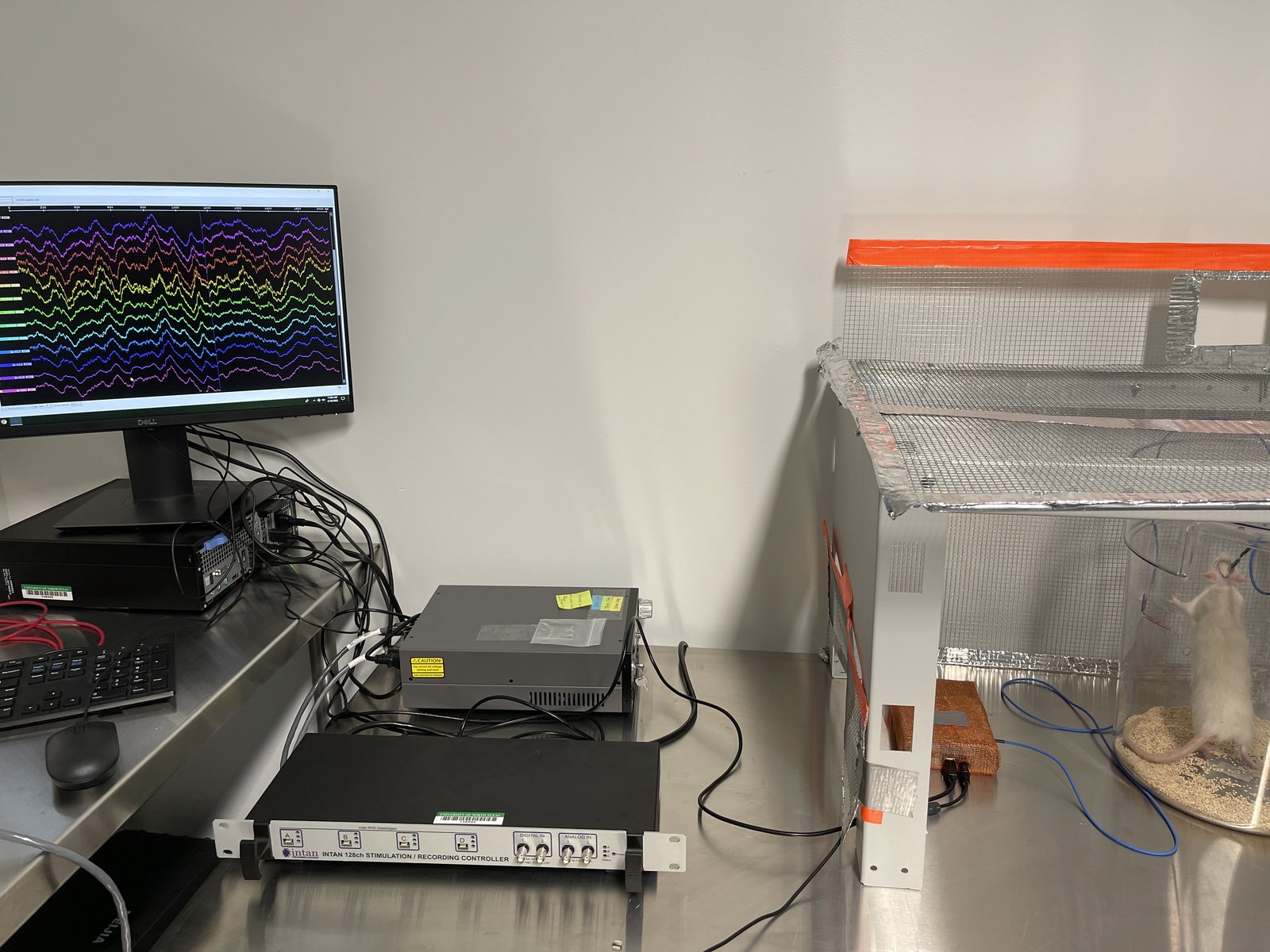
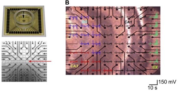
Multi-electrode array system
Multi-electrode array recordings
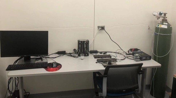
Preclinical recording setup
Preclinical neural recording platform
Computational analysis workstation
Large-scale signal analysis
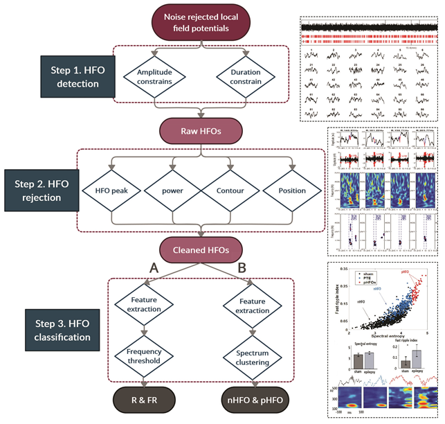
Spike-ripple and HFO waveform analysis
Automated event detection
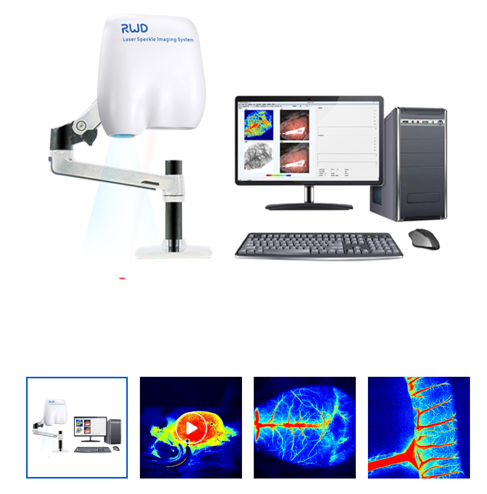
Laser speckle imaging system
Optical imaging systems
Laser speckle imaging lab setup
Brain hemodynamic imaging
Computational neuroscience analysis setup
Computational analysis workflows
Laboratory optical imaging equipment
Multimodal neuroengineering platform
Contribution 4

Computational and statistical methods for neuroscience.

Early work from the lab developed quantitative methods to improve reliability, image reconstruction, voxel classification, and interpretation in functional optical brain imaging. These methods enabled graph-theoretical analyses of brain networks and stronger study design for brain-behavior research.

This methodological foundation supports the lab's current multimodal integration across electrophysiology, MRI/fMRI, diffuse optical tomography, laser speckle imaging, and computational network science.

StatisticsReliability, reproducibility, and subgroup discovery methods.
ImagingAtlas-guided DOT, voxel classification, and optical neuroimaging.
IntegrationGraph theory and multimodal electrophysiology-imaging pipelines.

Representative publications

Choi et al. Recursive partitioning for subgroup identification in brain-behaviour correlation analysis. Pattern Analysis and Applications, 2020.

Li et al. Whole-cortical graphical networks at wakeful rest revealed by fNIRS. Neurophotonics, 2018.

Li et al. Automated voxel classification with atlas-guided diffuse optical tomography. Neurophotonics, 2016.

Li et al. Tutorial on intraclass correlation coefficients for fNIRS-based brain imaging. Journal of Biomedical Optics, 2015.

Lin et al. Atlas-guided volumetric diffuse optical tomography with GLM analysis. Human Brain Mapping, 2014.

Lin et al. Interleaved imaging of hemodynamics and blood flow in rat stroke models. NeuroImage, 2014.